Cancer research in Austin gets boost from $35,000 microscope
Scientists at The Hormel Institute know technology precedes important discoveries. Their latest acquisition to help further answers to cancer and other diseases is a state-of-the-art confocal microscope. The $350,000 Zeiss LSM 900 Confocal microscope provides a major upgrade to the existing confocal microscope and already researchers from 8 different labs have been trained to use the system.
“A lot of researchers use this instrument to continue research that they haven’t been able to do with our older system,” said Todd Schuster, instrument core facility manger. “Scientists are using online training to refine their skills and learn newer methods of imaging possible with this microscope. The new confocal is already being used by many different labs.”
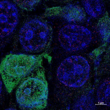
The Hormel Institute UMN now has 20 research sections, all dedicated to furthering answers to cancer and impacting other diseases as well. The confocal microscope – designed in Germany – uses a system of pinholes to remove out-of-focus light, resulting in sharper, higher quality images. Removing unfocused light allows researchers to focus on focal planes (the distance from the camera where the focus is sharpest) throughout the sample. By removing light that is out of focus, the scientists can distinguish between objects very close together. The ability to focus on focal planes within the sample helps scientists show different events happening in the same cell. Images from different levels of the sample can be obtained and put together to form a 3D image of the specimen being studied.
Upgrades to the detectors will result in higher resolution and better sensitivity. Imaging will be faster and the software has more features than the older system. Some of new software features include tiling where the software allows you to “stitch” together multiple fields of view to create one large image.
The Zeiss LSM 900 Confocal microscope replaced an older confocal microscope nearing the end of its lifespan and not functioning properly. Users were unable to obtain the images critically needed to further their research. The 600-pound instrument was shipped from Germany, arriving in four giant wooden crates.
“Having technology such as this is extremely important to further successful outcomes for the researchers at The Hormel Institute,” said Dr. Sergio Gradilone, Associate Professor and head of the Cancer Cell Biology and Translational Research lab.
“The new confocal enables precise, three-dimensional imaging, improved analyses of cellular organelles, and intracellular localization of proteins so we can study more accurately how cancer cells develop and metastasize, leading to more knowledge about how to prevent and control cancer.”
The technology was funded thanks to joint support from The Hormel Foundation and the Office of the Vice President for Research at University of Minnesota.
You Might Also Like